Updated by the Progyny Editorial Team. Reviewed by the Progyny Clinical Team — December 2025.
Structural problems in the uterus and fallopian tubes are one possible cause of infertility. You may have tests to look for these problems as part of a routine fertility evaluation. Or testing may be done if your doctor suspects you have an issue.
Diagnostic testing may be done to look for:
- Blocked fallopian tubes, which make it impossible for sperm to reach an egg for fertilization. A blockage can also prevent eggs from moving from the ovary to the uterus. (If a fertilized egg can’t travel to the uterus, it can lead to a life-threatening condition called an ectopic pregnancy.) Fallopian tubes may be blocked from pelvic inflammatory disease, scar tissue, endometriosis, or fibroids.
- Problems with the uterus, which can prevent an embryo from implanting in the endometrium (lining of the uterus). Uterine problems may include fibroids, polyps, scar tissue, and problems that can happen when the uterus is developing (the most common being a problem called uterine septum).
There are a few tests your doctor may recommend, each with benefits and drawbacks. Be sure to discuss your options together and ask any questions you have.
Saline infusion sonohysterogram (SIS)
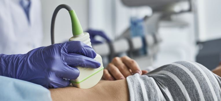
A saline infusion sonohysterogram (SIS) is a transvaginal ultrasound that is done while your uterus is filled with saline solution, to better see the inside of the uterus.
- What it detects: Issues in the uterus and endometrium, such as endometrial polyps, fibroids, or uterine scars.
- How it works: Saline solution is passed through the cervix to fill the uterus while a transvaginal ultrasound is done. This shows a more detailed picture of the uterus and endometrium than an ultrasound without saline.
- Benefits: A quick procedure (less than 10 minutes) that provides important information about issues that could prevent an embryo from implanting in the uterus.
- Drawbacks: Does not provide any information about the fallopian tubes.
Hysterosalpingogram (HSG)
A hysterosalpingogram is an x-ray that uses contrast dye to look at the fallopian tubes and uterus.
- What it detects: Blockages in the fallopian tubes and some problems in the uterus, such as uterine fibroids, polyps, scar tissue, uterine septum, or other problems.
- How it works: Contrast dye is carefully pumped into your uterus to fill the uterus. An x-ray will take pictures of the dye flowing through the uterus. If there is no fallopian tube blockage, the dye can pass through the fallopian tubes and into your body (where it’s safely absorbed). If the dye cannot pass, there is a blockage.
- Benefits: Provides information about the fallopian tubes, including where a blockage is located in the fallopian tubes, as well as some information about the uterus.
- Drawbacks: Involves a low dose of radiation, can be uncomfortable, and does not provide as much information about the uterus as an SIS.
Sonohysterosalpingogram (sono-HSG)
A sono-HSG is a newer method that does not use an x-ray. It provides information about the fallopian tubes and the uterus using saline solution and a transvaginal ultrasound.
- What it detects: Blockages in the fallopian tubes (including an issue called hydrosalpinx, where fluid builds up and blocks the fallopian tube) and problems in the uterus, such as uterine fibroids, polyps, scar tissue, uterine septum, or other problems.
- How it works: Saline solution, along with air, is passed through the cervix to fill the uterus. A transvaginal ultrasound will show the saline with air bubbles flowing through the uterus. If there is no fallopian blockage, the saline and air bubbles can pass through the fallopian tubes and into your body (where it is safely absorbed). If the saline and air bubbles cannot pass, there is a blockage.
- Benefits: Unlike HSG, there is no exposure to radiation and the test can be done in your fertility doctor’s office.
- Drawbacks: Not as well-studied as the HSG, not as widely available, and not as effective at evaluating the fallopian tubes.
Hysteroscopy
A hysteroscopy allows your provider to look inside the uterus. The uterus is filled with fluid to better see the inside, and a hysteroscope (a narrow, lighted camera in the shape of a tube) allows the provider to see any problems. A hysteroscopy is done to diagnose problems of the uterus, often including unusual bleeding.
- What it detects: Issues on the inside of the uterus or the endometrium. These may include polyps, fibroids, scar tissue, or problems related to how the uterus developed. Sometimes a hysteroscopy is done with other tools to take a tissue sample (biopsy) or treat problems (such as removing polyps). Your doctor would describe why the procedure may be needed in your case — it has many uses.
- How it works: Often, medicine is used to help you relax, or anesthesia may be used. A hysteroscope is passed through the cervix, and a liquid is sent into the uterus to help the doctor see more clearly. If needed, specialized tools can be inserted to perform surgical treatments.
- Benefits: This is a common, safe diagnostic procedure that can be performed along with surgery if needed in a single session.
- Drawbacks: Any procedure has risks. Hysteroscopy has a small risk of bleeding, infection, reactions to anesthesia, and other problems. Your doctor will describe these risks to you.
If you have questions about the tests your doctor has recommended for you, it’s always OK to ask them. Your Progyny Care Advocate is also here to help you understand your options and prepare for your conversations with your doctor.
Disclaimer: The information provided by Progyny is for educational purposes only and is not medical advice. Always consult a qualified healthcare provider for medical guidance.